The mammary gland is remarkably plastic due to mammary epithelial cells and their companion stem cells
The female human breast is a uniquely and cyclically plastic endocrine organ
The mammary gland is one of the most dynamic tissues in the human body. Unlike many organs that are largely established during embryogenesis, much of the mammary gland develops after birth, particularly at puberty and during pregnancy. The mammary gland's cyclical nature of development and regression throughout much of the lifespan is unusual amongst human endocrine organs, which usually develop embryologically and then perform consistently throughout life.
Researchers propose that this exceptionally dynamic or plastic nature of the female human breast has a downside: it makes the breast more vulnerable to malignant change than most other tissues in the body.
There are three main cell types which together create the highly dynamic architecture of the breast, enabling the cycles of growth, lactation, and regression. These are the two different cell-types of the mammary epithelium, and the stem or progenitor cells.
Mammary epithelial cells
The mammary gland epithelium has two layers
-
Luminal epithelial cells, which compromise the inner layer, facing the alveolar lumen, and comprising both secretory cells (lactocytes) and ductal cells. There are two types of luminal epithelial cells
-
Ductal luminal cells: Found in ducts, involved in transporting milk.
-
Alveolar luminal cells (lactocytes): Differentiate during pregnancy to produce milk components with secretory activation.
-
-
Myoepithelial cells form the outer layer of the epithelium in ducts and alveoli, between luminal cells and the basement membrane.They surround the luminal cells, sitting between them and the basement membrane. Myoepithelial cells have two functions. They
-
Are contractile cells that squeeze alveoli and ducts during milk ejection (“let-down”), under oxytocin stimulation.
-
Provide structural support, maintaining alveolus or duct shape
-
have a protective role, acting as a barrier against stromal invasion (by malignancy) and have tumour-suppressive properties.
-
Both luminal and myoepithelial cells arise from the mammary epithelium during embryonic development. They share a common progenitor pool in the basal layer of the epithelium.
Mammary epithelial cells act as 'non-professional' immune cells
Mammary epithelial cells are not passive secretory cells. They are multifunctional, immune-like participants in mammary gland physiology.
-
Mammary epithelial cells have been shown to participate in macrophage-like, phagocytic 'clearing up' activity of apoptotic cells and milk residue, during involution or apoptotic events in lactation (such as breast inflammation). They behave as 'non-professional' immune cells in other ways too, including
-
Pro-inflammatory cytokine and chemokine production, which recruit leukocytes when infection or stress is detected.
-
Production of anti-inflammatory signals after engulfing apoptotic cells, promoting immune tolerance and controlled tissue remodeling.
These actions have the following biological significance:
-
By clearing apoptotic cells themselves, MECs minimize the need for massive immune cell influx during involution, which reduces tissue damage.
-
MEC phagocytic activity helps maintain milk quality during lactation by removing dead cells and microbial contaminants.
-
By producing cytokines and antimicrobial peptides, MECs form part of the first line of defense in the breast against bacterial oveergrowth.
Mammary epithelial cells are highly plastic
Depending on physiological stage, MECs can exist as:
-
Quiescent cells (in non-pregnant gland).
-
Proliferative and differentiating cells (during pregnancy).
-
Secretory cells (during lactation, producing milk).
-
Apoptotic and cleared cells (during involution, when lactation ceases).
Mammary stem cells
The remarkable plasticity of the mammary gland is driven by the two different populations of stem and progenitor cells found within the bilayer of the mammary epithelium, which maintain tissue homeostasis and remodeling through a woman's reproductive life, enabling cycles of expansion, milk production, and regression.
1. Basal epithelial or stem-like cells
These basal epithelial or stem-like cells are grouped with myoepithelial cells in the basal layer. They
-
Include progenitor cells capable of giving rise to both luminal and myoepithelial lineages.
-
Are important for breast gland regeneration during pregnancy cycles.
2. Luminal stem cells
Distinct luminal progenitor cells have been identified, nestled amongst the lactocytes. These cells are thought to be responsible for generating both ductal luminal cells and the alveolar luminal cells (lactocytes) as they expand massively during pregnancy to enable milk secretion.
-
These luminal progenitor cells have been form colonies in culture and can generate alveolar-like structures in three-dimensional assays.
-
Luminal progenitors are of great clinical interest they are believed to be the cell of origin for several types of breast cancer, particularly basal-like and triple-negative breast cancers.
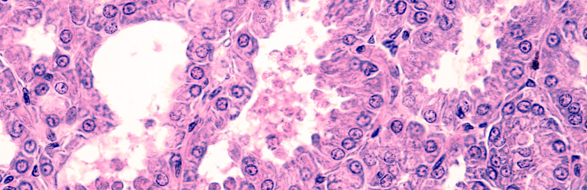
In the histological image at the top of this page, you can see long fingers projecting from the lactocytes which line the not-very-full alveolus in the centre, on its upper right margin. This shows milk fat globules are being pinched off from the lactocyte cell walls (parturition).
References
Monks J, Henson PM, Fadok VA, et al. (2005). Epithelial cells as phagocytes: apoptotic cell engulfment by mammary epithelial cells and mammary gland involution. J Cell Biol, 171(3): 431–441.
Atabai K, Werb Z. (2005). Mammary epithelial cells as nonprofessional phagocytes. J Mammary Gland Biol Neoplasia, 10(2): 163–170.
Rudland PS, et al. (2005). Mammary epithelial cells as multifunctional immune-like cells. J Mammary Gland Biol Neoplasia, 10(2): 119–136.
Thomas EC, Wlliams TM, Hartmann PE. Lactation and mother's milk: recent advances in understanding. Infant. 2010;6(3):86-90.
The photo at the top of the page is of tissue from a lactating mammary gland, demonstrating the lactocytes' apical activity.