The anatomy of the frenulum under your baby's tongue (known as the lingual frenulum)
The frenulum under your baby's tongue (or the lingual frenulum) is not a discrete band of connective tissue
Your baby's soft, moist, beautiful, sensing and sensitive baby-tongue is suspended in, and stabilized by, a pristine floor of mouth fascia. Quite marvellously, the fascia or connective tissue which creates the floor of your baby's (found under the surface layer of the mucosa) stabilises your baby's tongue while still allowing the tongue to be mobile. You can find out about your baby's tongue here.
New Zealand paediatric Ear Nose and Throat Surgeon Dr Nikki Mills, through painstaking anatomic dissections, discovered that the floor-of-mouth fascia is like a connective tissue sling, which suspends the tongue and the floor of mouth structures inside the bony arc of the jaw or mandible. Dr Mills discovered that the lingual frenulum is not a discrete 'band', the way we'd all been saying.
There is nothing in the cell composition of your baby's floor of mouth fascia to suggest there is a different composition or tightness or elasticity of the fascia (or frenulum) at the point where the tongue connects to the floor of mouth, and nothing to justify describing the frenulum as a band or discrete structure.
Dr Mills has devoted years to parsing out the functional anatomy of the infant suck and swallow. She likes to tell audiences - and we can hear the awe in her voice - that the tongue is an incredible organ. A strange muscle that doesn't connect to a bone, unlike every other muscle! A muscle that contracts and changes shape without changing volume, which makes it a 'muscular hydrostat'!
In the same way that Professor Donna Geddes's work on the functional anatomy of the lactating breast has re-written the textbooks, Nikki's painstaking work dissecting the floor of mouth oral connective tissues has also meant that anatomic text books need to be changed.
Three types of tissue can be found in your baby's lingual frenulum
Dr Mills discovered that there are three main kinds of tissue which can make up a lingual frenulum.
-
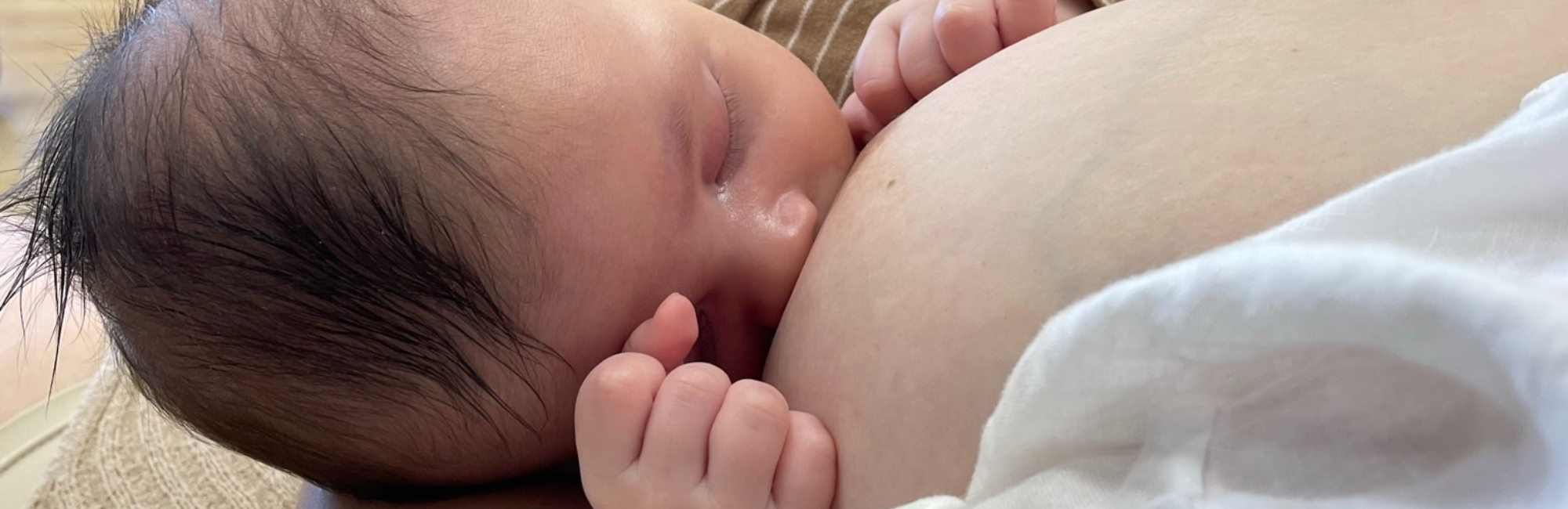
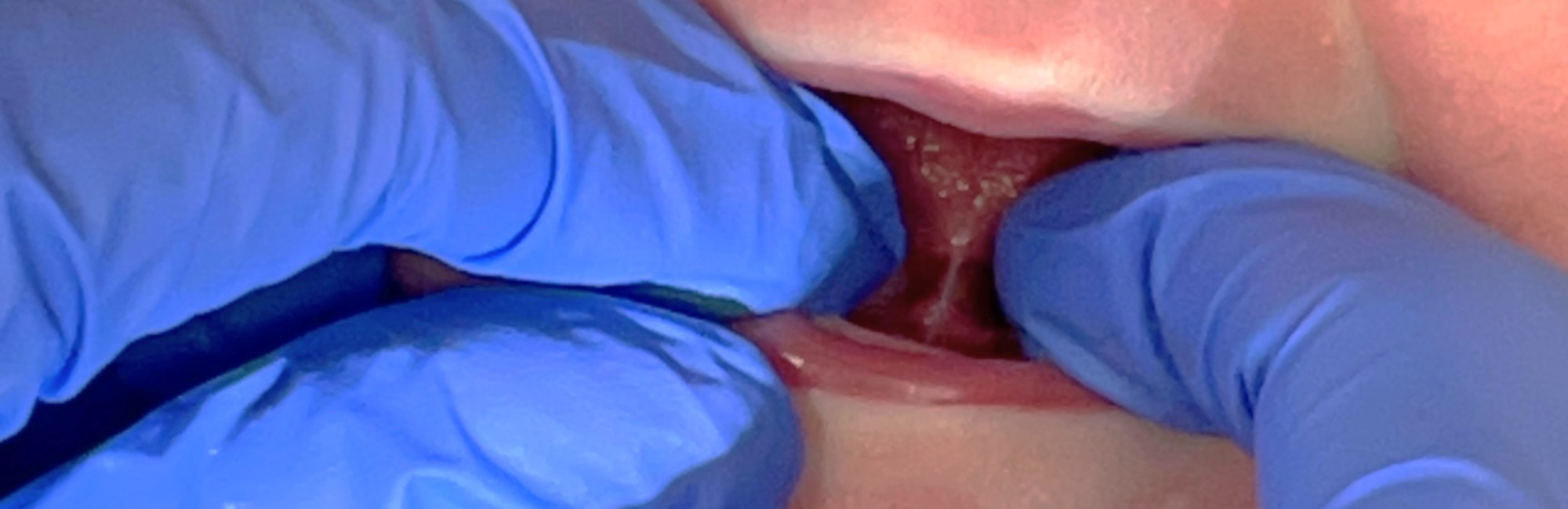
The floor of mouth mucosa often pulls up tent-like, under tension, when the tongue is lifted up. You can see an illustration of this transparent membrane in the photo at the top of this article.
-
Sometimes the floor of mouth fascia is also caught and pulled up tent-like too, under and inside the mucosa, when the tongue is lifted.
-
Sometimes some genioglossus muscle fibres are caught up, along with the floor of mouth fascia and the mucosa, as the tongue is lifted.

This is the diagram that Dr Mills published in 2019, illustrating her discoveries. Figure 1 shows how people have been wrongly thinking of the frenulum has a band of connective tissue (green) under the mucosa (red). Figure 2 shows the actual anatomy of the frenulum. Figure 2A shows how the mucosa (red) sits on top of the floor of mouth fascia (green) when the tongue is relaxed and down. Figure 2B shows how in many transparent frenulae, the mucosa pulls up under the tongue like a tent when the tongue is lifted. The floor of mouth fascia isn't visible in the frenulum. Figure 2C shows how in some frenulae, the floor of mouth fascia, with its connective tissue, follows the mucosa up into a tent-like structure when the tongue is lifted. Figure 2D shows how in other frenulae, the floor of mouth fascia and the genioglossus muscle both pull up into a tent-like structure under the mucosa when the tongue is lifted.The baby in the picture at the top of the page has a more prominent frenulum, which might contain some floor of mouth fascia under the mucosa as it tents when the tongue is lifted. This baby presented with breastfeeding problems which were attributed to tongue-tie, but the problems resolved with a gestalt intervention.
Right now, Dr Mill's work is still often misinterpreted as showing how tightly the floor of mouth fascia can be pulled up under the tongue, and that this fascia needs to be stretched and softened. This isn't true! Each is a normal variation, whether it's just the mucosa, or the mucosa plus fascia, or the mucosa, fascia and genioglossus muscle which are pulled up when the tongue is lifted.
Ongoing unhelpful pathologising of the lingual frenulum and other oral connective tissues has shifted the breastfeeding baby's care from the hands of the surgeon who performs a frenotomy, to the traditional bodyworker.
-
You can find out about bodywork and me here.
-
You can find out what science tells us about traditional bodywork here.
-
You can find out about evolutionary bodywork here.
-
You can find out about how the baby's tongue moves in breastfeeding here.
-
You can find out about evolutionary bodywork here.
There is no anatomic or functional basis to the idea of a posterior tongue-tie
I first met Dr Mills on a podium in Auckland, at the New Zealand Lactation Consultant's Association annual conference. That was when she told me that she regularly saw, using fiberoptic laryngoscopy, that the base of the infant tongue moves forwards and backwards a little with suckling. Later that night, she generously took me back to her home, since she felt it would be easier for me to get to the airport for my pre-dawn flight from there. We became friends.
Dr Mills has confirmed that the concept of 'posterior tongue-tie' misunderstands the nature of the frenulum, and has no place as a diagnosis. There is no fascial connection between the posterior or base of the tongue, and the floor of mouth fascia.
-
You can find out about the diagnosis of posterior tongue-tie here.
-
You can find a short history of the diagnosis of posterior tongue-tie here.
Selected references
-
Mills N, Pransky SM, Geddes DT, Mirjalili SA. What is a tongue tie? Defining the anatomy of the in-situ lingual frenulum. Clinical Anatomy. 2019:doi:10.1002/ca.23343.
-
Mills N, Keough N, Geddes DT, Pransky S. Defining the anatomy of the neonatal lingual frenulum. Clinical Anatomy. 2019;32:824-835.
-
Mills N, Geddes DT, Amirapu S, Mirjalili SA. Understanding the lingual frenulum: histological structure, tissue composition, and implications for tongue tie surgery. International Journal of Otolaryngology. 2020(1820978.):doi: 10.1155/2020/1820978.
-
Mills N, Lydon A-M, Davies-Payne D, Keesing M, Mirjalili SA, Geddes DT. Imaging the breastfeeding swallow: pilot study utilizing real-time MRI. Laryngoscope Investigative Otolaryngology. 2020;5:572-579.
-
Mills N, Keesing M, Geddes DT, Mirjalili SA. Flexible endoscopic evaluation of swallowing in breastfeeding infants with laryngomalacia: observed clinical and endoscopic changes with alteration of infant positioning at the breast. Annals of Otology, Rhinology and Larygology. 2020:doi:10.117/00034894220965636.