White spots on the nipple during lactation: management
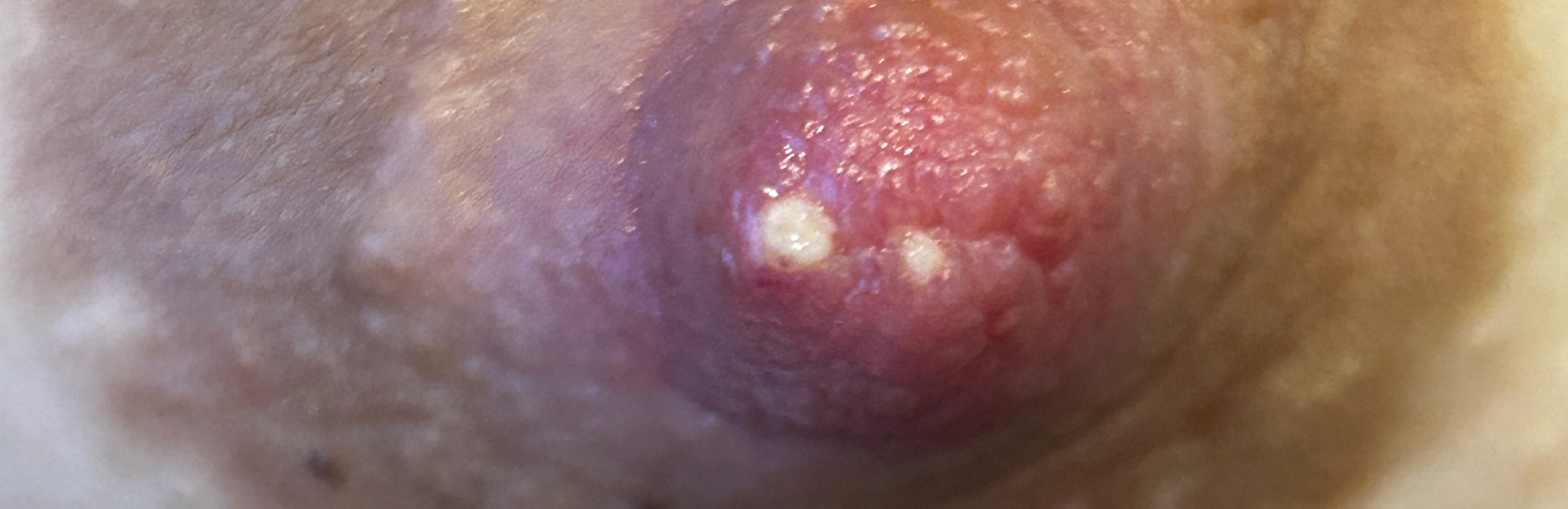
1. Management of a hyperkeratotic spot of nipple during lactation (common)
The gestalt method is currently the only fit and hold intervention which offers an evidence-based biomechanical model for eliminating conflicting intra-oral vectors of force during milk removal.1-3 Using the NDC Guidelines, management of a hyperkeratotic white spot requires
-
Distribution of mechanical load over a larger area of nipple and areola surface, by eliminating conflicting intra-oral vectors of force during suckling or mechanical milk removal, in order to eliminate repetitive mechanical micro-trauma
-
Application of steroid cream like mometasone twice daily for the first day, then daily for a week. Whilst there is no evidence to support this approach, it has a sensible biological rationale: to temporarily suppress the inflammatory response, at the same time as repetitive microtrauma of breast tissue drag is being addressed.
Occlusive dressing or ointment should be avoided, because moisture associated skin damage increases the vulnerability of the nipple epithelium to micro-trauma
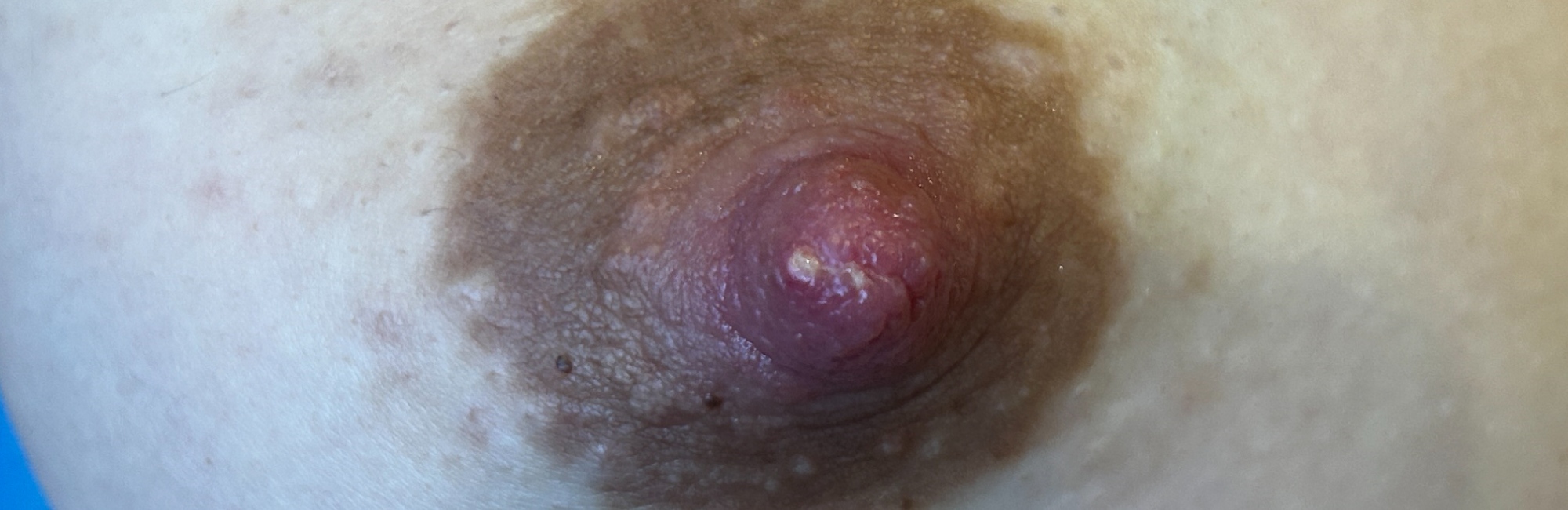
2. Management of a milk blister (rare)
If you are confident that the woman has a milk blister, an epidermal rool which is covering a milk duct
-
A beveled needle may be used to lift the epithelial roof. Often, there is immediate leakage of milk once the bleb is unroofed.
-
Advise the woman to breastfeed her infant from that breast as frequently as possible, including immediately after deroofing. Short frequent episodes of milk flow through the orifice, once breast tissue drag is eliminated, may prevent the roof re-sealing over the duct.
Advise women not to rub at the nipple with a cloth or fingernail or attempt to deroof the bleb themselves (which they are often advised to do in the shower), as hyperkeratosis can result from self-treatment, particularly if it is repeated. If a milk blister persists, a steroid cream may suppress the inflammatory response which causes the roof to form.
Light therapy or photobiomodulation is successfully used for treatment of sublingual salivary gland mucoceles, which are retention cysts similarly caused by an epithelial roof at the duct orifice. Research is required to investigate whether laser has a role in the case of recurrent milk blisters, but from the perspective of biological plausibility, it may have a role.
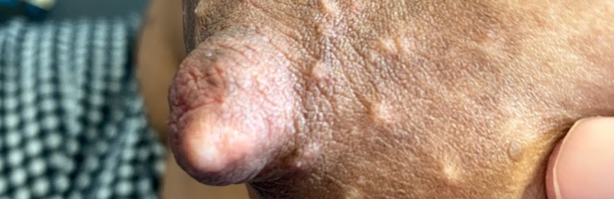
3. Management of a milium on the nipple (occasional)
A milium requires no intervention.
4. Management of a larger epidermal inclusion cyst (occasional)
An epidermal inclusion cyst requires complete surgical excision of the cyst with its walls intact, which prevents reoccurrence. Excision is best accomplished when the lesion is not acutely inflamed. If a woman has an epidermal inclusion cyst of her nipple-areolar complex evident during pregnancy, which may be large enough to interfere with breastfeeding, then it needs to be removed with plenty of time left in the pregnancy for complete healing. Ducts may be severed during removal, which could predispose to a breast inflammation during lactation. However, ducts may also be occluded if the inclusion cyst is left intact, which could also predispose to a breast inflammation during lactation. The decision about the best time to intervene - and whether or not surgical intervention is necessary - will require clinical judgement, taking into account that woman's unique context and preferences.
References
- Douglas PS, Geddes DB. Practice-based interpretation of ultrasound studies leads the way to less pharmaceutical and surgical intervention for breastfeeding babies and more effective clinical support. Midwifery 2018;58:145–55.
- Douglas PS, Keogh R. Gestalt breastfeeding: helping mothers and infants optimise positional stability and intra-oral breast tissue volume for effective, pain-free milk transfer. Journal of Human Lactation 2017;33(3):509–18.
- Douglas PS, Perrella SL, Geddes DT. A brief gestalt intervention changes ultrasound measures of tongue movement during breastfeeding: case series. BMC Pregnancy and Childbirth 2022;22(94):https://doi.org/10.1186/s12884-021-04363-7.