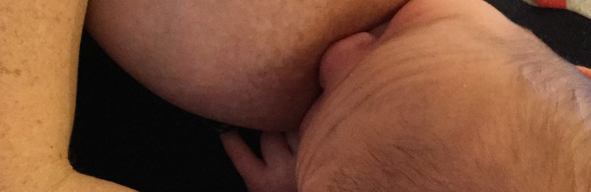
Nipple wounds (cracks, blisters, ulcers) result when the epidermis fractures as a result of focussed high mechanical stretching loads
Nipple pain with visible damage results when excessively high intra-oral mechanical loads fracture the epithelium
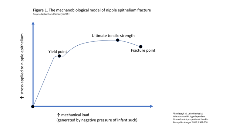
If epithelium can no longer adapt to the mechanical strain of stretching, bending and shearing forces, and the desmosomes have locked but the stretching force continues to increase, epithelium ruptures at the ‘fracture point’.1 The more humid the environment, resulting in more tissue hydration, the longer the cracks.2
In the NDC mechanobiological model of lactation-related nipple pain, the weakest part of the nipple-areolar complex epithelium, or the part placed under the most constant and severe elastic tension in the baby’s mouth, will be the first part of the epidermis to break apart.
This results in visible trauma, including cracks, grazes, and ulcers, with associated pain and inflammation.
The figure below illustrates the mechanobiological model of nipple epithelium yield (when tight junctions lock) and fracture (when the epidermis breaks). This graph adapted from Pawlaczyk 2013. 5
What causes cracks?
Skin cracks start in the stratum corneum along the plane of maximum shear stress, as desmosomes rupture. At the microscopic level, cracks in the superficial cell layers travel along intercellular junctions. Fracture of the corneocytes or keratinocyte cells themselves is uncommon.
-
Cracks propagate along the topographical crevice or 'canyon' features of the epithelium of the nipple face, at the bottom of an epidermal 'canyon'.2
-
Cracks are also often located at the base of the nipple, which depending on the vectors of mechanical force may be where the plane of maximum shear stress occurred.
-
It is sometimes (but not always) possible to determine the direction of the intra-oral conflicting force from the location of a crack.
- For instance, if the crack is at 6 o’clock at the base of the nipple adjacent to the areola, the infant may be suckling from the breast at a height above the natural gravity-induced breast fall, causing the mechanical strain of upward tension on the intra-oral nipple and breast tissue.3-5
-
When the stratum corneum, ruptures, it becomes ineffective at regulating water loss and preventing external pathogens from infecting underlying living tissue. Fortunately, the exposed underlying tissue has its own protective mechanisms which immediately come into play.
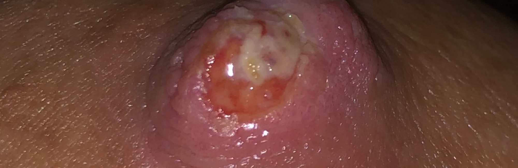
What causes grazes or ulcers?
Wider areas of epithelial damage (ulcers) develop and spread as women continue placing their baby on their breast or expressing milk mechanically in the context of existing epithelial damage.
What causes blisters?
Blisters result when horizontal shearing forces cause partial fracture and inflammatory serum collects in a pocket of fluid between layers of skin. Bruising results from vascular damage and haemorrhage.
References
- Pawlaczyk M, Lelonkiewicz M, Wieczorowski M. Age-dependent biomechanical properties of the skin. Postep Der Alergol 2013;5:302-06.
- Liu X, Cleary J, German GK. The global mechanical properties and multi-scale failure mechanics of heterogeneous human stratum corneum. Acta Biometerialia 2016;43:78-87.
- Douglas PS, Geddes DB. Practice-based interpretation of ultrasound studies leads the way to less pharmaceutical and surgical intervention for breastfeeding babies and more effective clinical support. Midwifery 2018;58:145–55.
- Douglas PS, Keogh R. Gestalt breastfeeding: helping mothers and infants optimise positional stability and intra-oral breast tissue volume for effective, pain-free milk transfer. Journal of Human Lactation 2017;33(3):509–18.
- Douglas PS, Perrella SL, Geddes DT. A brief gestalt intervention changes ultrasound measures of tongue movement during breastfeeding: case series. BMC Pregnancy and Childbirth 2022;22(94):https://doi.org/10.1186/s12884-021-04363-7.