The lactating nipple-areolar complex has unique protections and unique risks
How does the lactating nipple-areolar complex adapt to the unique stressors of lactation and breastfeeding?
There are four main protective systems which interact in the skin of your nipple-areolar complex during lactation to maintain skin health and homeostasis.
The following key protective systems interact in the skin of the lactating nipple-areolar complex to maintain health and homeostasis.
-
Host immune system
-
Skin and milk microbiome
-
Adaptation to repeated mechanical loads. You can find out about this here.
-
Wound-healing inflammation. You can find out about the mechanisms of wound healing here.
Table 2. The lactating nipple-areolar complex is characterised by unique protective factors and also exposed to unique risks, relative to other parts of human skin
| Unique NAC risk | Unique NAC protective factors |
|---|---|
| Areolar sweat and mammary glands secrete more moisture than many other skin sites, increasing the risk of moisture associated skin damage. | |
| Female nipple dermis has dense concentration of nociceptors (pain perception) | If we know how to respond to even mild signals of pain, we can protect the nipple from damage. |
| Exposure to repetitive and frequent mechanical load from the negative pressure of suckling, applied perhaps two to four hours in total during a 24-hour period. | A. The nipple face has deep epithelial crevices and ridges, which enhance epithelial elasticity and distribute mechanical loads. B. Keratinocytes adapt to repetitive mechanical loads by 1. Elongation 2. Changing orientation to align with direction of mechanical forces, and 3. Proliferation. |
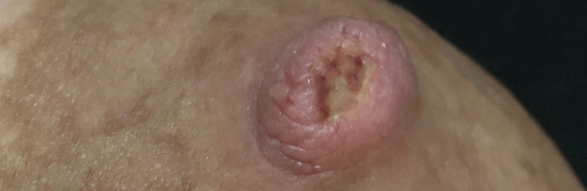
| Exposure to excessively high stretching and deforming forces caused by conflicting intra-oral vectors of force during suckling or mechanical milk removal results in epithelial and stromal inflammation, epithelial damage, and nociceptor stimulation. | Nipples are richly vascularised, resulting in unusually rapid transport of immune and wound-healing factors. A normal layer of nipple epidermis (not exposed to repeated micro-trauma and environmental humidity) may recover from damage in around three days, depending on depth of injury, compared to 7-10 days for damaged epidermis elsewhere on the skin. When there is exudate and necrotic eschar, cyclic mechanical stress under negative pressure exposes a nipple wound to repetitive debridement. Wound exudate is protective. Scab formation is part of the healing process. The infant mouth is an ideal cleansing and debriding application due to antimicrobial and immunoprotective effects of infant saliva, human milk, and milk and oral microbiome. (Box 4) |
| Lack of a subcutaneous layer exposes nipple stroma to the shearing and stretching effects of excessively high mechanical forces during suckling or mechanical milk removal, resulting in microvascular haemorrhages, an inflammatory cascade, and pain. | NDC proposes that lack of subcutaneous tissue has three evolutionary advantages. 1.Vacuum acts directly upon superficial lactiferous ducts without an added cushioning layer. 2.Ductal dilation is optimized without the added intra-oral volume of subcutaneous tissue. 3.The nipple can achieve a firm shape. |
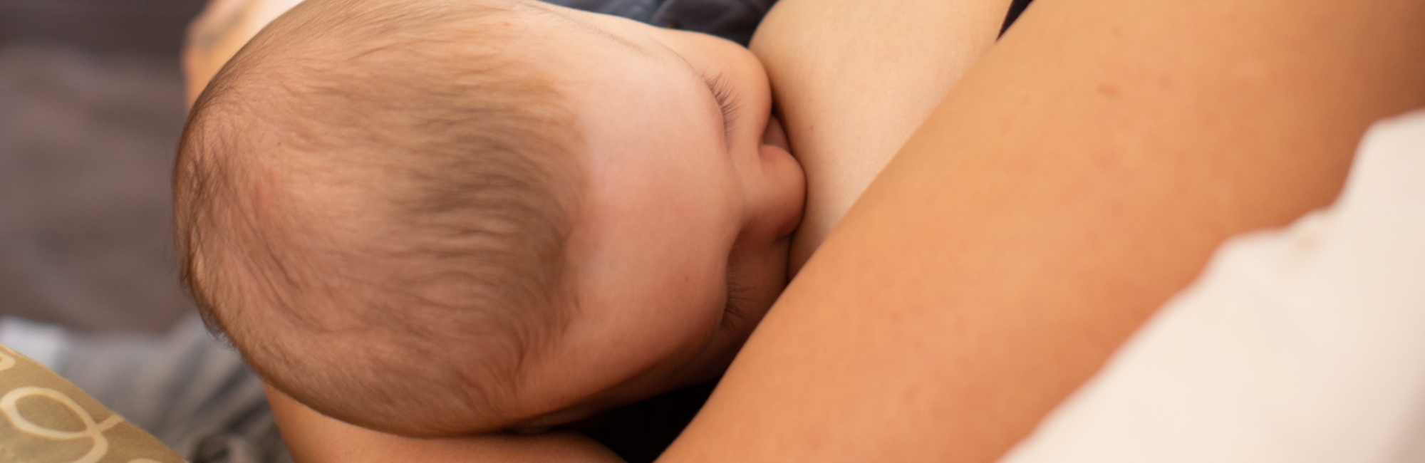
| Epithelial breaks due to exposure to excessively high mechanical forces are vulnerable to microbial overgrowth due to loss of the protective epithelial barrier. | Saliva, the infant oral microbiome, nipple skin, and breastmilk including the milk microbiome interact to form the infant's oral mucosal immune system. A. Nipples are frequently bathed in human milk which contains myriad interacting immunoprotective factors including the microbiome, metabolome, immune cells, and exosomes. B. Infant saliva contains multiple interacting bactericidal, fungicidal, and immunoprotective factors. The growth of micro-organisms is inhibited by breastmilk alone and more so when infant saliva is mixed with breast milk, with synergistic bactericidal effects. |
| Bras and breast pads form an occlusive dressing (that is, do not act as semi-permeable membranes). Occlusive dressings result in increased temperature, increased carbon dioxide and decreased oxygen levels, increased humidity, and increased acidity. These changes predispose to nipple epithelial overhydration and Moisture Associated Skin Damage, which increases risk of epithelial fracture. Breast pads also absorb moisture (milk and sweat). This further predisposes to nipple epithelial overhydration and Moisture Associated Skin Damage, which increases risk of epithelial fracture. | |
| Many milk ducts travel just a millimetre or two deep to the fascia of the areola and breast skin. There is no subcutaneous tissue, which means that milk ducts are very susceptible to compression with even light pressure, much like touching a vein on the back of your hand. |
References
- Esfahani AM, Rosenbohm J, Reddy K. Tissue regeneration from mechanical stretching of cell-cell adhesion. Tissue Engineering and Regenerative Medicine 2019;25(11):doi:10.1089/ten.tec.2019.0098.
- Douglas PS, Geddes DB. Practice-based interpretation of ultrasound studies leads the way to less pharmaceutical and surgical intervention for breastfeeding babies and more effective clinical support. Midwifery 2018;58:145–55.
- Douglas PS, Keogh R. Gestalt breastfeeding: helping mothers and infants optimise positional stability and intra-oral breast tissue volume for effective, pain-free milk transfer. Journal of Human Lactation 2017;33(3):509–18.
- Douglas PS, Perrella SL, Geddes DT. A brief gestalt intervention changes ultrasound measures of tongue movement during breastfeeding: case series. BMC Pregnancy and Childbirth 2022;22(94):https://doi.org/10.1186/s12884-021-04363-7.
- Douglas PS. Re-thinking benign inflammation of the lactating breast: a mechanobiological model. Women's Health 2022;18:https://doi.org/10.1177/17455065221075907.
- Sweeney EL, Al-Shehri SS, Cowley DM, et al. The effect of breastmilk and saliva combinations on the in vitro growth of oral pathogenic and commensal microorganisms. Scientific Reports 2018;8:15112.
- Pan L, Zhang X, Gao Q. Effects and mechanisms of histatins as novel skin wound-healing agents. Journal of Tissue Viability 2021;30:190-95.
- Hajishengallis G, Russell MW. Innate humoral defense factors. Mucosal Immunity 2015:251-70.