How nipple-areolar complex wounds heal during lactation
Mechanisms of nipple-areolar complex wound healing during lactation
Acute wound healing is characterised by four overlapping phases: haemostasis, inflammation, proliferation, and remodelling.1 Bacterial presence is required for wound regeneration and healing.
Haemeostasis
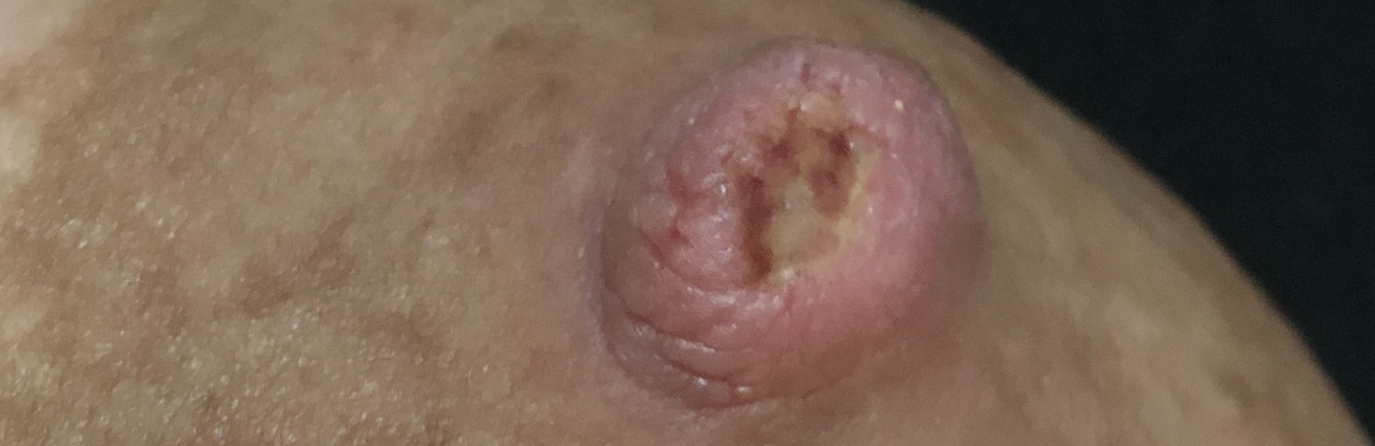
Once excessive stretching forces result in rupture of nipple epithelial desmosomes, the dermis too may tear, accompanied by microscopic or visible bleeding. Epithelial rupture with or without dermal injury triggers a cascade of inflammatory signals, as an efficient and evolutionary process of wound healing begins.
Platelets are carried by the blood stream to the site of damage, combining with blood cells and fibrin to create a clot, which provides a scaffold for incoming inflammatory cells. Neutrophils are immediately recruited as a first line of defence against unfriendly bacteria or overgrowth of particular species of bacteria which exists in the microbiome.
Inflammation
A cascade of signals from broken desmosomes, microvascular trauma, and platelets continue to trigger the inflammatory response, which dilates vasculature and increases endothelial permeability, resulting in exudate. This thin, clear or cloudy moisture is remarkable for its healing properties.
-
Exudate contains proteins such as fibrinogen and fibrin and is rich in electrolytes, leukocytes, and proteases, and also protective bioactive molecules, including various growth factors, cytokines, and hormones.
-
Exudate promotes keratinocyte proliferation and fibroblast growth.
-
Exudate neutrophils and later, macrophages, clean the wound of dead or dying tissue.
-
-
The moisture of the exudate allows cells in the wound area communicate with each other, ensuring coordinated healing. (Acute wound exudate is very different to the exudate of chronic wounds like diabetic or vascular wounds, which are not relevant to nipple damage, even when persistent. Chronic wounds have high levels of proteases.)1
The moisture, warmth and nutrients of the wound area alter the microbiome, creating opportunities for colonisation by new microorganisms or for expansion of existing microorganisms. To heal, diverse populations of bacteria, fungi and other micro-organisms dynamically interact, modulating each other’s growth, regulated by the homeostatic control of the host’s immune system.2
As the clot hardens and dehydrates, an eschar scab forms. The scab may be yellowish, orange, or brown. It is dehydrated and composed of dead cells in a fibrin mesh which seals the breach in the epidermis and acts as a carapace.
An epithelial blister similarly acts like bubble wrap, protecting the wound. Both keep other micro-organisms out and allow keratinocytes and broken blood vessels underneath to heal.
Proliferation
Synthesis of collagen by fibrocytes generates a new granulation tissue matrix. Angiogenesis (or blood vessel proliferation) facilitates rich vascularisation of the granulation tissue and extracellular matrix deposition, providing oxygen and nutrients necessary for tissue synthesis. Oxygen also kills harmful anaerobic bacteria.
Remodelling
Stem cells continuously renew keratinocytes, which proliferate and migrate upward from the basal layer to fill the wound defect. In all phases of wound healing, keratinocytes interact with fibroblasts, endothelial cells, immune cells, growth factors and cytokines in a coordinated manner.
Epidermal cells from sweat glands and the wound periphery also migrate to re-epithelialize the wound. Tissue remodelling and re-epithelisation of the wound leads to reconstitution of the physical barrier of the nipple skin.1
References
-
Rodrigues M, Kosaric N, Bonham CA, Gurtner GC. Wound healing: a cellular perspective. Physiological Reviews. 2019;99(1):665-706.
-
Drago F, Gariazzo L, Cioni M. The microbiome and its relevance in complex wounds. European Journal of Dermatology. 2019;29(1):6-13.