Milk production is irregular throughout the breast but the transport mechanism is robust
Volumes of milk synthesis are highly variable in successfully breastfeeding women
Studies investigating mechanical milk removal and also 24 hour test weighing of breastfed infants have elucidated the range of rates of milk synthesis typical for the breasts of successfully breastfeeding women.1, 2
When exclusive breastfeeding is successful, the amount of milk secreted by a woman’s breasts rapidly stabilises from day 11 postpartum at an average of 788 gm daily. The range in successful breastfeeding is highly variable (600 gm – 1220 gm) depending on the mother-infant pair.2, 14
The NDC mechanobiological model of breast inflammation hypothesises that
-
Downregulation of milk secretion throughout the course of lactation is predominantly mechanobiological
-
Upregulation of milk secretion is facilitated by sensory stimulation of suckling and the hormonal effects of more frequent milk removal on stem cells.
Human stem cells are derived from the maternal haematopoietic system and remodel the breast when development of lactocytes and myoepithelial cells is required. Stem cells are located in the myoepithelial and epithelial layers of alveoli and ducts, presumably not only in Type 3 and 4 but also in under-developed Type 1 and 2 lobules.
Prolactin stimulates not only milk synthesis but also cell proliferation, which is theorised to be the mechanism which utilises the stem cells to result in regeneration and differentiation of the lactating epithelium and dynamic maintenance and turnover of the secretory tissue during the course of the lactation.15-19
Rates of milk synthesis differ randomly between different parts of the glandular tissue
-
Between 20%-100% of the glandular tissue of lactating women is comprised of highly productive lobules, which are collections of alveoli emptying into a common ductule. These are characterised as Type 3 or Type 4 lobules. Significantly, 20% of lactating women have less than 60% of Type 3 and 4 lobules.
-
The remainder of the glandular tissue is undifferentiated or less mature lobules, labelled as Types 1 and 2, from which lower amounts of milk are secreted.
In a series of pioneering ultrasound imaging studies, Geddes et al (also Ramsay et al) have demonstrated that about two-thirds of alveoli and their lobules within a lactating breast are located within a 3 cm radius of the nipple, and that lactiferous ducts travel back from nipple orifices to the alveoli in densely interlaced branching patterns.
Ducts ranging from 0.1 - 10 mm in diameter at rest have been identified under the areola with ultrasound, but even narrower ductules run to the alveoli. Because there is little subcutaneous tissue under the dermis of the nipple-areolar-complex, ducts are often just 1-2 millimetre beneath the surface, and are highly compressible with even very light external touch, much like veins on the back of the hand.4, 7, 8
It is likely that many ductules rest for a time between feeds in an occluded or closed down state, much like the 50% of lymphatic vessels which are collapsed and quiescent in the lactating breast.3 Ducts may also gradually fill with milk which is constantly secreted by the lactocytes and which flows out along pressure gradients between feeds. The ducts of each breast may fill with up to 30 mls of milk, transferred to the infant by vacuum application of suckling prior to oxytocin activation, but there are no ‘lactiferous sinuses’ which store milk.4
Frequency of milk removal in the first few days of life sets the amount of milk that can be produced down the track
The capacity to generate more alveoli and ducts throughout the course of lactation appears to be limited by base-line numbers of prolactin receptors, set in the first hours and days of life.
-
In 140 healthy term Japanese newborns, 7-11 opportunities for milk removal in the first 24 hours postpartum was associated with increased 24-hour milk production, decreased weight loss, and decreased serum bilirubin by days 5-7.20
-
In 358 healthy term Nigerian newborns who breastfed about 13 times in the first 24 hours showed improved weight gain and lower serum bilirubin levels by day 7 compared to those who fed less frequently.21
-
Rate of milk production at two weeks post-birth in 98 healthy term infants in the United States correlated with frequency of milk removal and predicted the rate of milk production at 6 weeks.22
How does milk leave the lactating breast?
The lactating mammary gland is a highly dynamic and adaptive environment. Milk ejection, for example, is not machine-like and precise. As in all biological systems, there is a great deal of asynchrony and variability, though milk ejection remains, overall, well-coordinated and robust.
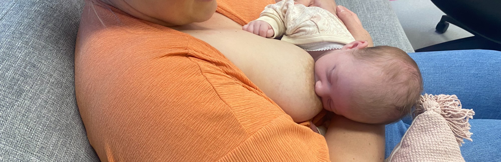
There are three ways milk leaves the lactating breast. Milk is removed from the breasts by
-
The intermittent negative mechanical pressure of suckling, and
-
The intermittent positive mechanical pressure of milk ejection. Most milk leaves the breast when these two mechanisms work together in tandem.
-
Sometimes the breasts leak milk in the absence of suckling, due to positive pressure of milk ejection alone.
Gravity does affect milk transfer
Lying back does not decrease the amount of milk transferred from a lactating breast. Gravity does not affect milk transfer - though a semi-reclined position may impact positively upon fit and hold, and help a baby dial down at the breast because of improved positional stability.
Selected references
Please note that the referencing in this module is still under development. Comprehensive citations are found in the three research publications which the breast inflammation module is built (Douglas 2022 mechanobiological mode; Douglas 2022 classification, prevention, management; Douglas 2023)
Douglas P. Re-thinking benign inflammation of the lactating breast: a mechanobiological model. Women's Health. 2022;18:17455065221075907.
Douglas PS. Re-thinking benign inflammation of the lactating breast: classification, prevention, and management. Women's Health. 2022;18:17455057221091349.
Douglas PS. Does the Academy of Breastfeeding Medicine Clinical Protocol #36 'The Mastitis Spectrum' promote overtreatment and risk worsened outcomes for breastfeeding families? Commentary. International Breastfeeding Journal. 2023;18:Article no. 51 https://doi.org/10.1186/s13006-13023-00588-13008.
- Kent JC, Gardner H, Lai C-T, Hartmann PE, Murray K, Rea A, et al. Hourly breast expression to estimate the rate of synthesis of milk and fat. Nutrients. 2018;10:1144.
- Kent JC, Gardner H, Geddes DT. Breastmilk production in the first 4 weeks after birth of term infants. Nutrients. 2016;8(756):doi:10.3390/nu8120756.
- Jindal S, Narasimhan J, Vorges VF, Schedin P. Characterization of weaning-induced breast involution in women: implications for young women's breast cancer. Breast Cancer. 2020;6(55):https://doi.org/10.1038/s41523-020-00196-3.
- Ramsay DT, Kent JC, Hartmann RA, Hartmann PE. Anatomy of the lactating human breast redefined with ultrasound imaging. Journal of Anatomy. 2005;206:525-34.
- Murase M, Mizuno K, Nishida Y, Mizuno H. Comparison of creamatocrit and protein concentration in each mammary lobe of the same breast: does the milk composition of each mammary lobe differ in the same breast? Breastfeeding Medicine. 2009;4(4):189-95.
- Gardner H, Kent JC, Hartmann PE, Geddes DT. Asynchronous milk ejection in human lactating breast: case series. Journal of Human Lactation. 2015;31(2254-259).
- Ramsay DT, Kent JC, Owens RA, Hartmann PE. Ultrasound imaging of milk ejection in the breast of lactating women. Pediatics. 2004;113:361-7.
- Geddes DT. The use of ultrasound to identify milk ejection in women - tips and pitfalls. International Breastfeeding Journal. 2009;4(5):doi:10.1186/746-4385-4-5.
- Stewart TA, Hughes K, Stevenson AJ, Marino N, Ju AL, Morehead M, et al. Mammary mechanobiology - investigating roles for mechanically activated ion channels in lactation and involution. Journal of Cell Science. 2021;134:doi:10.124/jcs.248849.
- Mortazavi N, Hassiotou F, Geddes DT, Hassanipour F. Mathematical modeling of mammary ducts in lactating human females. Journal of Biomechanical Engineering. 2015;137(7):071009.
- Prime DK, Geddes DT, Hepworth AR, Trengove NJ, Hartmann PE. Comparison of the patterns of milk ejection during repeated breast expression sessions in women. Breastfeeding Medicine. 2011;6(4):183-90.
- Gardner H, Kent JC, Prime DK, Lai C-T, Hartmann PE, Geddes DT. Milk ejection patterns remain consistent during the first and second lactations. American Journal of Human Biology. 2017;29:e22960.
- Gardner H, Kent JC, Lai CT, Geddes DT. Comparison of maternal milk ejection characteristics during pumping using infant-derived and 2-phase vacuum patterns. International Breastfeeding Journal. 2019;14(47):https://doi.org/10.1186/s13006-019-0237-6.
- Kent JC, Mitoulas LR, Cregan MD, Ramsay DT, Doherty DA, Hartmann PE. Volume and frequency of breastfeedings and fat content of breast milk throughout the day. Pediatics. 2006;117(3):e387-e95.
- Kersin SG, Ozek E. Breast milk stem cells: are they magic bullets in neonatology? Turkish Archives of Pediatrics. 2021:doi:10.5152/TurkArchPediatr.2021.21006.
- Witkowska-Zimny M, Kaminska-El-Hassan E. Cells of human milk. Cellular and Molecular Biology Letters. 2017;22(11):doi:101186/s11658-017-0042-4.
- Ninkina N, Kukharsky M, Hewitt MV. Stem cells in human breast milk. Human Cell. 2019;32:223-30.
- Li S, Zhang L, Zhou Q. Characerization of stem cells and immune cells in preterm and term mother's milk. Journal of Human Lactation. 2019;35(3):528-34.
- Hassitou F, Hartmann PE. At the dawn of a new discovery: the potential of breast milk stem cells. Advances in Nutrition. 2014;5:770-8.
- Yamauchi Y, Yamanouchi I. Breast-feeding frequency during the first 24 hours after birth in full-time neonates. Pediatrics. 1990;86(2):171-5.
- Okechukwu A, A O. Exclusive breastfeeding frequency during the first seven days of life in term neonates. Nigerian Postgraduate Medicine Journal. 2006;13(4):309-12.
- Hill PD, Aldag JC, Chatterton RT, Zinaman M. Primary and secondary mediators' influence on milk output in lactating mothers of preterm and term infants. Journal of Human Lactation. 2005;21(2):138-50.
- Hassiotou F, Geddes DT. Anatomy of the human mammary gland: current status of knowledge. Clinical Anatomy. 2013;26:29-48.
- Oliver G, Kipnis J, J RG, Harvey NL. The lymphatic vasculature in the 21st century: novel functional roles in homeostasis and disease. Cell. 2020;182:270-96.
- Schwager S, Detmar M. Inflammation and lymphatic function. Frontiers in Immunology. 2019;10:doi:10.3389/fimmu.2019.00308.
- Steele MM, Lund AW. Afferent lymphatic transport and peripheral tissue immunity. The Journal of Immunology. 2021;206:264-72.
- Moore JE, Bertram CD. Lymphatic system flows. Annual Review of Fluid Mechanics. 2018;50:459-82.
-fotor-20230723183658.jpg)